Hip pain or the presence of a limp when walking in children can be particularly important to investigate, in most cases the underlying cause can be put down to a sporting or play incident and will resolve with appropriate treatment. However, in some less common conditions this limp or presence of pain in the hip and knee can be indicators of more serious pathology such as Perthes Disease, Slipped Upper Femoral Epiphysis (SUFE) or Hip dysplasia.
The hip joint functions in a ball and socket mechanism with the ball (head of femur) and socket (acetabulum) forming a stable joint. This joint continues to develop throughout childhood and If not formed correctly there is an increased likelihood of long term pain and limitations.
A muscle strain and tear in either the hip or knee can present similarly with pain and causing a limp therefore it is important to identify the correct source of pain. At Physica, our physiotherapists are trained to identify early signs and risk factors of these conditions such as leg length discrepancy, decreased hip range, worsening pain with activity, increased limping or antalgic gait and decreased muscle development that can indicate the need for an X-ray or further investigation. Early identification of conditions affecting this bony formation such as Perthes Disease, SUFE and hip dysplasia is very important and early treatment is associated with better long term outcomes.
Perthes Disease
Perthes disease is the interruption of blood flow (avascular necrosis) to the femoral head causing softening and damage to the bone. This affects the formation of a smooth and round ball shaped femoral head. It typically presents in children between the ages of 3-12 and is found more commonly in males.
An early indication of Perthes disease is the development of an insidious painless limp, generally followed by hip, groin, lateral thigh or knee pain that is worsened with time on the affected leg. An X-ray will confirm diagnosis by showing a flattening and possible fragmentation of the femoral head.
Treatment for Perthes focuses on helping the bone grow back into a more rounded shape that still fits into the socket of the hip joint. This will help the hip joint move normally and prevent hip problems in adulthood. This is where physiotherapy is important to help maintain strength , mobility and provide load management advice for the return to activities during the bone healing timeframe. The long-term prognosis for children with Perthes is good in most cases. After 18 months to 2 years of treatment, most children return to daily activities without major limitations.
Slipped Upper Femoral Epiphysis
A ‘Slipped Upper Femoral Epiphysis’ (also called a ‘SUFE’) is a condition where the growth plate (called the epiphyseal plate) at the top of the thigh bone is weak and the head of the femur slips downward and backward. The exact cause of this condition is not known, but there may be a link between increased weight and puberty hormones.
A ‘SUFE’ is not usually associated with an injury most often; it develops during periods of accelerated growth, shortly after the onset of puberty (mainly ages 10-16). The symptoms often develop slowly – over several months – and may seem like a pulled muscle in the hip, thigh or knee. Children may present with just knee pain that worsens to a limp or restricted range of movement in the hip.
It is important to get an early diagnosis and treatment before the slip gets worse and in 20% of cases children may need to have the unaffected side treated as well to prevent future slipping. Treatment depends on how severe the slip is. Most children need an operation to place metal screws into the head of the femur to for stability.
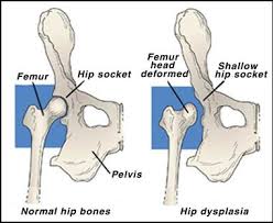
Hip dysplasia
Hip dysplasia is the abnormal development of the hip joint that can lead to subluxation or dislocations. It is commonly checked for in babies that have the following risk factors – breech birth, female, family history and can be identified through the use of specialised testing and Ultrasound imaging at 6 weeks. Hip dysplasia can be difficult to diagnose initially and in some cases, hip dislocations have developed in older babies even when the infant exam was normal and the ultrasound was normal at six weeks of age. The cause of these late developing cases is unknown however an increased risk factor can be swaddling tightly or extended periods of legs in a more straightened position and it is not picked up until weight bearing or walking stages.
In older children the term Hip dysplasia generally refers to the hip joint being the wrong shape, or that the hip socket is not in the correct position to completely cover and support the femoral head. This causes increased force, and abnormal wear on the cartilage and labrum. Hip Dysplasia can have a variety of presentations with more severe resulting in dislocations and requiring surgery to restore function to more mild that respond well to conservative management such as physiotherapy exercises to strengthen the hip and decrease joint load or a period of bracing and positioning advice to improve the formation of the joint. It is important to have this identified early by your physiotherapist and have treatment methods clearly outlined and explained to ensure optimal joint functioning.