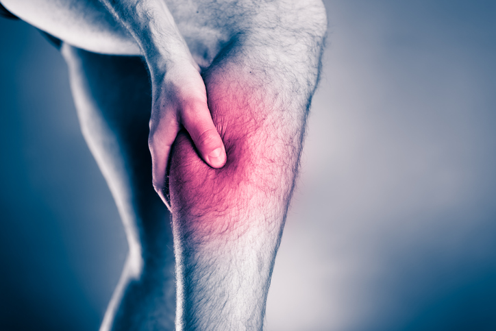
WHAT IS COMPARTMENT SYNDROME?
Compartment syndrome is a condition that involves an excessive pressure build up within a space in the body (Tiwari, Haq, Myint, & Hamilton, 2002). The increase in pressure can result from bleeding or swelling, which in turn causes pain to the region and can sometimes result in compression of local nerve and arteries (Shears & Porter, 2006).
There are two main types of compartment syndrome; acute and chronic. Acute compartment syndrome is often more serious and usually results in more extreme measures for treatment (Via, Olivia, Spoliti, & Maffulli, 2015), whereas chronic compartment syndrome is less threatening and associated with exercise (Tucker, 2010). The areas of the body most prone to compartment syndrome are in the extremities, specifically the lower leg and forearms, though cases are known to occur almost anywhere (Shears & Porter, 2006).
The most common causes for acute compartment syndrome are (Tiwari et al., 2002):
-
- Fractures (particularly the tibia)
- Surgery to repair fractures
- Crush injury
- Burns
- Prolonged compression of a limb
- Snake bite
- Infection
- Surgery to blood vessels (particularly the arm or leg)
- Extremely vigorous exercise (though this is rare for acute compartment syndrome)
Whereas the most common cause of chronic compartment syndrome is regular vigorous exercise, particularly running in young males (Tucker, 2010). It is also known as chronic exertional compartment syndrome and develops over days to months, whilst acute compartment syndrome results from a sudden incident usually involving trauma.
Chronic compartment syndrome can be viewed as a form of ‘overuse injury’ and can result in changes in load a shorter than ideal space of time (George & Hutchinson, 2012). This can vary enormously throughout the population as some present soon after changes and others can take months. Also, some changes to load and exercise can be quite drastic, whereas others can be subtle, such as changing the type of shoe you run in (George & Hutchinson, 2012).
ANATOMY
Regions of the human body are often organised into compartments. The areas most prone to compartment syndrome usually contain muscles and of course the accompanying nerve and arteries. The compartments are contained by fascial walls. Fascia is a type of soft tissue in the body that is comprised of fibrous connective tissue; it is utilised for rigid support and as such is less flexible than other soft tissue like muscle and therefore does not expand greatly.
The most common region for the development of compartment syndrome, the lower leg, contains four compartments:
- Anterior (front) compartment
- Lateral compartment
- Deep posterior (back) compartment
- Superficial posterior compartment (more commonly known as your calf region)
WHAT ARE THE SIGNS AND SYMPTOMS OF COMPARTMENT SYNDROME?
Common symptoms of acute compartment syndrome are (Garner, Taylor, Gausden, & Lyden, 2014):
- Pain
- Paraesthesia (numbness, tingling or burning)
- Paralysis or weakness of muscles in that region or below that region i.e. weakness to muscles in the foot as a result of compartment syndrome in the calf
Pain, tingling and numbness are often early signs. In later stages pain may be absent and weakness or paralysis of muscles may be noticed.
In cases of chronic compartment syndrome, pain is usually associated with or after exercise. In some cases, tingling or numbness is associated but rarely does it progress beyond this.
DO I HAVE COMPARTMENT SYNDROME?
Diagnosis of compartment syndrome is mostly made clinically. This involves understanding the recent history of the individual i.e. presence of recent trauma or incident, recent fracture or surgery.
Clinicians examination involves (Garner et al., 2014):
- Palpating and stretching the local area for pain
- Assessing for lack or diminished pulse in the area below
- Coldness to the region
- Assessment of strength
- Assessment of numbness
If acute compartment syndrome is suspected, referral to the Emergency Department for pressure testing is required. Pressure testing involves the measure of pressure within the compartment via a needle device (McQueen, Duckworth, Aitken, & Court-Brown, 2013). However, a negative pressure test does not exclude the diagnosis if the clinical presentation is compelling.
Tissue pressure (McQueen et al., 2013):
Normal pressure: 0-10 mmHg
Capillary blood flow at risk: 10-20 mmHg
Muscle and nerve fibres at risk of ischaemia: >30 mmHg
Diagnosis using pressure testing involves a Delta pressure, which is calculated simply by:
Delta pressure = diastolic blood pressure – intracompartment pressure (measured with device)
In the case of chronic compartment syndrome, the above assessment for diagnosis is usually unchanged (Bong, Polatsch, Jazrawi, & Rokito, 2005), however pressure testing, if required, can be performed by a surgeon at a later time as this is usually a non-emergency. Clinical testing is also likely to involve exercise to understand the threshold and severity of the symptoms (George & Hutchinson, 2012).
TREATMENT OF COMPARTMENT SYNDROME
For acute compartment syndrome a fasciotomy may be required. This is a surgical procedure involving fascia of a compartment being cut to relieve the pressure (Garner et al., 2014). The recovery time varies between cases, but most uncomplicated cases involve a 4-6-week recovery (Tiwari et al., 2002).
In less serious cases of acute compartment syndrome not requiring fasciotomy treatment involves, elevation of the limb to the level of the heart and removal of all constrictive dressings. Symptoms are monitored very closely during this period to ensure improvement is seen as a fasciotomy may still be required (Al-Dadah, Darrah, Cooper, Donell, & Patel, 2008).
Acute compartment syndrome is often an emergency and if left untreated can result in ischaemia (restriction in blood supply to tissue) leading to muscle and nerve cell death. Ultimately resulting in loss of function to the region and in some cases, gangrene and potential loss of limb or rhabdomyolysis (breakdown of skeletal muscle tissue) leading to kidney failure (Tiwari et al., 2002).
Chronic compartment syndrome is most commonly managed conservatively (Bong et al., 2005). The most effective form of treatment is load management. This usually involves careful planning with a physiotherapist regarding the type, intensity and amount of exercise that an individual can safely do without worsening the condition (Tucker, 2010). In many cases periods of rest or low-intensity activity may be required for symptoms to settle. Patients may also find relief and faster progression to normal activities with massage to the affected area (Winkes, van Eerten, & Scheltinga, 2020). Stretching based exercises have demonstrated a role in conservative management of this condition. In some cases, foot biomechanics during running or other activities also play a part, therefore foot orthotics may be prescribed for management (George & Hutchinson, 2012). Conservative treatment should continue for a period of 6-12 weeks before more aggressive treatment options are considered.
DIFFERENTIAL DIAGNOSIS
For acute compartment syndrome, other possible diagnoses may include (Garner et al., 2014):
- Deep vein thrombosis (DVT)
- Cellulitis
- Peripheral vascular injuries
For chronic compartment syndrome, the differential diagnosis depends on the region that is affected. Usually other overuse syndromes such as medial tibial stress syndrome (shin splints), stress fracture or nerve entrapment syndromes can be possible diagnoses (George & Hutchinson, 2012).
REFERENCES
Al-Dadah, O. Q., Darrah, C., Cooper, A., Donell, S. T., & Patel, A. D. (2008). Continuous compartment pressure monitoring vs. clinical monitoring in tibial diaphyseal fractures. Injury, 39(10), 1204-1209. doi:10.1016/j.injury.2008.03.029
Bong, M. R., Polatsch, D. B., Jazrawi, L. M., & Rokito, A. S. (2005). Chronic Exertional Compartment Syndrome Diagnosis and Management. Bulletin of Hospital for Joint Diseases, 62(3&4), 77-84.
Garner, M. R., Taylor, S. A., Gausden, E., & Lyden, J. P. (2014). Compartment syndrome: diagnosis, management, and unique concerns in the twenty-first century. HSS J, 10(2), 143-152. doi:10.1007/s11420-014-9386-8
George, C. A., & Hutchinson, M. R. (2012). Chronic exertional compartment syndrome. Clin Sports Med, 31(2), 307-319. doi:10.1016/j.csm.2011.09.013
McQueen, M. M., Duckworth, A. D., Aitken, S. A., & Court-Brown, C. M. (2013). The estimated sensitivity and specificity of compartment pressure monitoring for acute compartment syndrome. J Bone Joint Surg Am, 95(8), 673-677. doi:10.2106/JBJS.K.01731
Shears, E., & Porter, K. (2006). Acute compartment syndrome of the limb. Trauma, 8, 261-266.
Tiwari, A., Haq, A. I., Myint, F., & Hamilton, G. (2002). Acute compartment syndromes. British Journal of Surgery, 89, 397-412.
Tucker, A. K. (2010). Chronic exertional compartment syndrome of the leg. Curr Rev Musculoskelet Med, 3(1-4), 32-37. doi:10.1007/s12178-010-9065-4
Via, A. G., Olivia, F., Spoliti, M., & Maffulli, N. (2015). Acute compartment syndrome. Muscles, Ligaments and Tendons Journal, 5(1), 18-22.
Winkes, M., van Eerten, P., & Scheltinga, M. (2020). Deep posterior chronic exertional compartment syndrome as a cause of leg pain. Unfallchirurg, 123(Suppl 1), 3-7. doi:10.1007/s00113-019-0665-1